
Understanding MRI Knee Anatomy: A Simple Guide for Beginners
Introduction
Understanding MRI Knee anatomy can feel confusing at first, especially if you are new to medical terms. But learning the basics is easier than it seems. An MRI gives a clear picture of the inside of your knee, helping doctors find problems early. This guide will break everything down into simple words so you can feel confident when your doctor explains your results.
MRI scans use strong magnets and radio waves to create detailed images. These images show the bones, muscles, cartilage, and ligaments inside your knee. When you know what these parts do, it becomes easier to understand why knee pain happens and how doctors decide the right treatment for you.
What an MRI Shows Inside the Knee
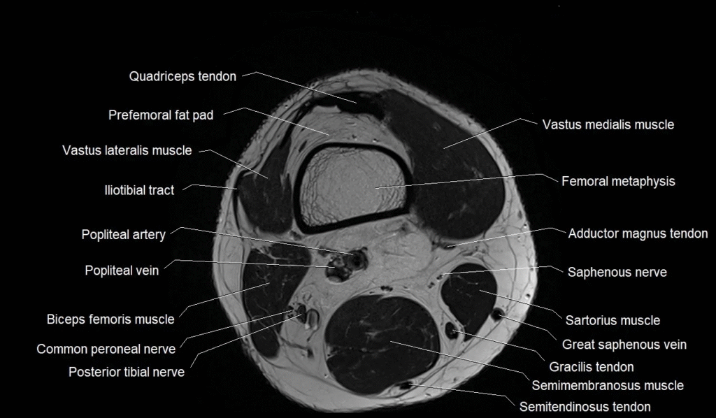
An MRI takes pictures from different angles, showing every important structure in the knee. This includes bones like the femur, tibia, and patella. It also shows soft tissues such as ligaments and cartilage. These details make MRI Knee anatomy helpful for spotting injuries that regular X-rays may miss.
The scan is especially useful when you have pain that does not improve or when you feel your knee popping, locking, or giving out. By viewing the knee clearly, doctors can see if something is torn, swollen, or damaged.
Cartilage and Meniscus in MRI Knee Anatomy
The cartilage in your knee acts like a cushion, helping the bones move smoothly. On an MRI, healthy cartilage looks smooth and even. When it becomes worn out or damaged, you may feel pain or stiffness. Cartilage problems are common in athletes and older adults.
The meniscus is another important part of MRI Knee anatomy. It is shaped like a rubber wedge and helps absorb shock. A meniscus tear can happen when you twist your knee suddenly. On an MRI, doctors can easily see if the meniscus is torn or inflamed, which helps them choose the best treatment plan.
Ligaments and Their Role in Knee Stability
Ligaments keep the knee stable and prevent it from moving in the wrong direction. The knee has four main ligaments: ACL, PCL, MCL, and LCL. These parts are clearly seen on MRI images. If any of them are torn, the knee may feel weak or unstable.
When doctors review MRI Knee anatomy, they look at the shape and condition of each ligament. A partial tear may only need rest and therapy, while a full tear might require surgery. Understanding these differences helps patients know what to expect.
Bones and Joint Alignment in MRI Knee Anatomy
The bones shown in an MRI help doctors see if the knee joint is aligned properly. Misalignment can cause pain, swelling, or long-term problems. The kneecap, or patella, must sit in the right position to move smoothly. If it shifts out of place, it may cause discomfort during walking or bending.
MRI scans also show bone bruises or small fractures that might be missed on other tests. This makes MRI Knee anatomy a powerful tool for finding hidden injuries and planning proper treatment.
Common Problems Seen on an MRI
People often get an MRI when they have knee pain after sports, exercise, or accidents. Some common problems include sprains, tears, arthritis, and swelling inside the joint. These issues show up clearly on MRI images, helping doctors understand the exact cause of the pain.
In many cases, the MRI helps avoid unnecessary treatments. When doctors see the real problem inside the knee, they can suggest the right medicine, therapy, or surgery based on your condition.
Conclusion
Learning about MRI Knee anatomy is helpful for anyone dealing with knee pain or recovering from an injury. Understanding what the MRI shows can make you feel more in control of your health. It also makes it easier to discuss results with your doctor and understand the next steps in your treatment.
With this simple guide, beginners can now understand the basic parts of the knee and how an MRI helps find problems quickly. Knowing these details brings confidence and clarity during your medical journey.